

Layer It Up: Studying Renaissance Paintings with Multispectral Spectroscopy
With custom hardware and software, the National Gallery uses three different methods of imaging spectroscopy in order to optimize the preservation of paintings for coming generations.
The Feast of the Gods is one of the masterworks of Italian Renaissance painter Giovanni Bellini. Created with oil paint on canvas, the scene depicts a group of mythological gods in a forest clearing with a shrub-lined hill in the background, and is based on the writings of the ancient Roman poet Ovid.
But The Feast of the Gods is not all that it seems.
Bellini finished the painting in the year 1514. He died in 1516, leaving the painting in the collection of Alfonso, I d’Este, the Duke of Ferrara, who had originally commissioned the work. After Bellini’s death, The Feast of the Gods was altered several times, first by painter Dosso Dossi and later by Titian.
A rudimentary X-ray analysis of the painting in the 1950s showed how The Feast of The Gods had been altered after Bellini’s death. But the technology at the time allowed little insight into how the painting was changed, and by whom. It would take modern, non-destructive imaging techniques and software at the National Gallery of Art in Washington, D.C. to get to the truth behind the painting’s evolution.
Three Methods of Spectroscopy Unveil Artistic History
A team at the National Gallery of Art headed by John K. Delaney, the senior imaging scientist in the scientific research department of the conservation division at the museum, uses a unique blend of hardware and software to analyze paintings.
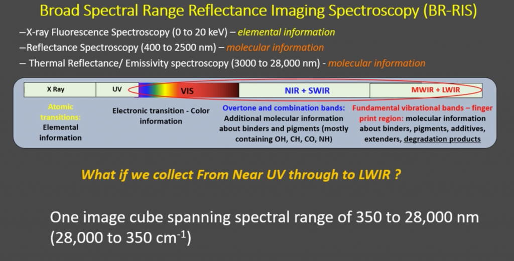
The imaging techniques are all a form of spectroscopy, which is the branch of science concerned with the investigation and measurement of spectra produced when matter interacts with, or emits, electromagnetic radiation. Basically, by studying light and other forms of radiation waves, scientists can tell what kind of matter objects are made from. Spectroscopy is a fundamental tool in the fields of chemistry, astronomy and physics, and has famously been used to study the chemical composition of distant stars and exoplanets.
Old paintings are exceedingly vulnerable. Conservators need to be able to study the paintings without damaging or destroying them. Paintings need to be stored and presented with optimal lighting conditions, at the right temperature. That means that conservators cannot beam excess light or drastically increase the temperature of the paintings when they are imaging them.
Chemical analysis of a painting often requires a physical sample scraped from the canvas, which is not a viable method when studying the full canvas of paintings that are often 500 years old or older.
To achieve the goals of studying the paintings without harming them, Delaney’s team employs three different forms of imaging spectroscopy:
- Diffuse Reflectance Imaging Spectroscopy (RIS)
- Molecular Fluorescence Imaging Spectroscopy (FIS)
- Elemental Fluorescence (X-ray Fluorescence) Imaging Spectroscopy (XRF-IS)
Diffuse reflectance, also known as hyperspectral imaging, “occurs when direct light impinges on the surface of a material and is partially reflected and transmitted.” In essence, light is bounced off a surface at a specific angle and its reflection is then measured. According to a 1998 paper published by Jonathan P. Blitz, diffuse reflectance is, “used if a sample is not shiny or amenable to conventional transmission spectroscopy.”
Blitz further explains:
“To explain what diffuse reflectance is, consider a white wall illuminated by sunlight. The wall appears equally bright regardless of whether you stand directly in front of it, or at any other angle. The surface of the wall reflects the incident sunlight at angles independent of the angle of incidence. In other words, the surface of the wall scatters the incoming radiation. Ideal diffuse reflection requires that the angular distribution of the reflected radiation be independent of the angle of incidence.”
Fluorescence spectroscopy, “uses a beam of light that excites the electrons in molecules of certain compounds and causes them to emit light,” which is then directed to an instrument for measurement and identification. The method allows researchers to study the luminescence—the light emission from a molecule—to determine what kind of matter it is made of based on the emission spectrum.
X-ray fluorescence works when a painting sample is irradiated by high-energy X-rays or gamma rays. According to ColourLex, a research firm that specializes in paintings, “The energy of the x-rays is sufficient to expel electrons from the inner shells (close to the atomic nucleus) in an atom. Electrons from outer shells fall subsequently into the holes left by the expelled electrons and emit x-rays in the process. As every atom has its own particular structure of its electron shells, the energy of the emitted x-rays is characteristic for each element and can be used for its identification.”
In this way, x-ray fluorescence can be used to determine the chemical characteristics of pigments used in the painting. For instance, “sodium can be assigned to ultramarine, mercury to vermilion and copper to either malachite, azurite or verdigris,” according to ColourLex.
Chemical analysis of a painting often requires a physical sample scraped from the canvas, which is not a viable method when studying the full canvas of paintings that are often 500 years old or older.
Mapping Pigments and Binders
Renaissance-era painters had many tricks to create a variety of colored paints, plus binding agents to make sure the paint stuck to the canvas. For instance, a certain pigment of blue may be mixed with egg yolk to serve as both a binding agent and to turn the blue into green.
The goal is to analyze pigments and binders without having to take physical samples from the painting, while adhering to the physical conditions in which the painting is kept. Delaney’s team explains:
“The conservation of a painting requires an understanding of the materials (pigments and binders) and construction methods used by the artist. There are numerous analytical methods used today that provide a wealth of detailed information about the materials used as well as the stratigraphy of the paint layers. These methods, such as SEM–EDS, high-performance liquid chromatography, and transmission-mode mid-IR spectroscopy, all require dispersed samples (scraping from the paint surface) or paint cross-sections. While advances in technology and the development of new protocols by conservation scientists have minimized the sample size needed, the taking of samples is still required. Even though these methods provide the most detailed chemical information about the materials used, they cannot be used across the entire painted surface because the number of samples allowed is limited.
The three different methods of spectroscopy used at the National Gallery of Art allow the team to analyze the makeup of paintings along the full wavelength spectrum, from near infrared all the way through the long wave.
“We use these different imaging modalities to address questions that have been posed by conservators and art historians, questions like what pigments were used in painting this composition?” Delaney said in a presentation published by SPIE in April 2020. “Where are the pigments found on the painting? And other such questions like, can you tell us more about the working methods of a given artist? Did they start with a preparatory drawing, or did they use a painted sketch? And probably the most fun part is to look for hidden changes in the paintings. That is, did the artist paint over a prior composition?”
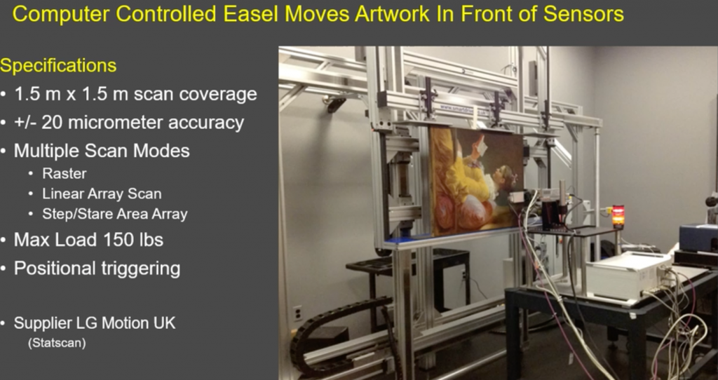
The equipment at the National Gallery of Art includes a Macro-XRF scanner, two hyperspectral cameras, and a VNIR camera used collect fluorescence emissions. Each camera or scanner is placed on a table and the paintings are set on a moving easel, which allows the gallery to easily change the equipment heads for different kinds of scans. Camera selection becomes quite important, as the conservators have to avoid excess light levels in their measurements to help protect the artworks. Low levels of reflected, fluorescence or X-Ray radiation require very sensitive imaging systems, typically used for scientific applications.
Each imaging modality used returns a what is called a data cube, which is a multi-dimensional array of values of the information that, in this case, the characteristics of the pigments and bonding agents of a given painting. By mining the information in these data cubes with proprietary algorithms (developed in cooperation with George Washington University and Rochester Institute of Technology), researchers can create a chemical map of the painting.
The Evolution of a The Feast of the Gods
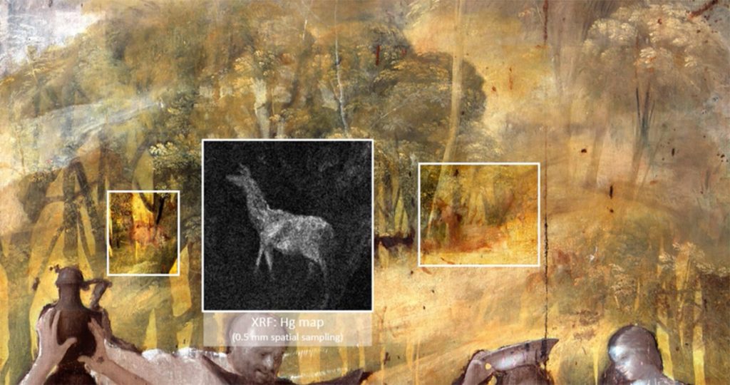
When the National Gallery of Art applied these modalities to The Feast of the Gods, what it found was that Bellini never painted the shrub-laden hill that dominates the left side background of the painting. Bellini’s original scene had the gods taking a picnic in front of a stand of trees. The hillside was later added by Dosso, and later refined by Titian.
Initial X-ray and infrared scans were done on the painting in the late 1980s that showed these changes. With X-ray matched with short wave infrared (SWIR) from the new scans, Delaney’s team was able to uncover more details, such as that the trees in Bellini’s version extended all the way to the top of the canvas. With this detail, a conservator reproduced what Bellini’s original painting looked like.
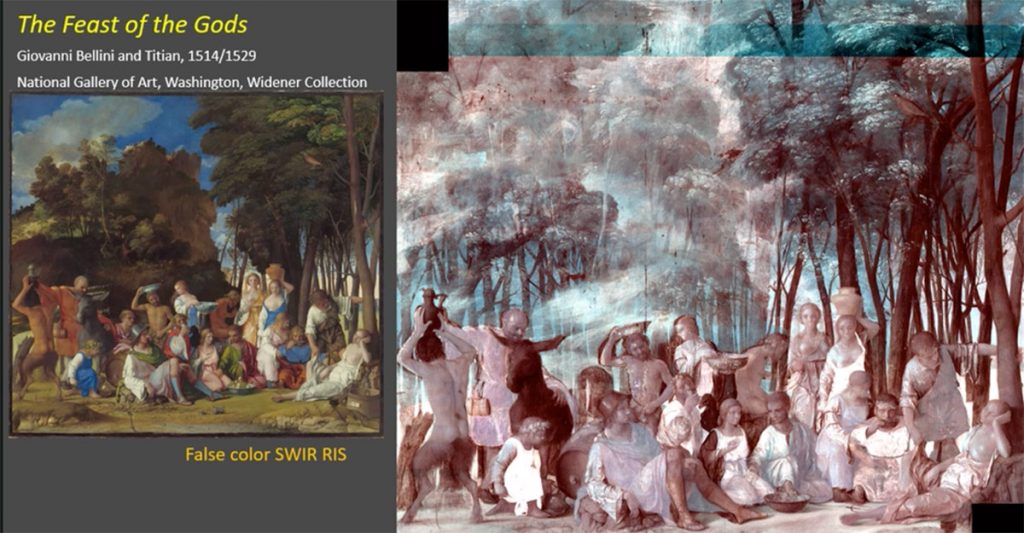
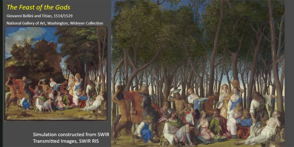
Before Titian finished the painting in 1529, Dosso added the hillside with a few of the artist’s signature bushy trees which showed up when analyzing the SWIR hyperspectral data cube. What surprised researchers most though were three deer in Dosso’s version that are not present in the finished work. By making use of the elemental imaging modality, researchers were able to identify the mercury used in the vermilion pigment used to paint the deer.
The National Gallery of Art has used these imaging modalities on a variety of paintings, helping uncover the techniques used by some of the great painters of the last thousand years. Researchers have scanned works from luminaries such as Van Gogh, Leonardo da Vinci, and Fragonard to tell stories about how works of art evolved, and come to conclusions about the artists’ motivations.
And while a peek behind the curtain of history is useful and important, these imaging modalities serve practical purposes as well. Conservators use the information obtained from the imaging spectroscopy to help preserve the paintings in as close to their original form as possible.
“Historical paintings are considered to be among the most precious cultural heritage artifacts and have been the subject of intensive studies for decades,” researchers from University of Antwerp wrote in 2014. “Scientific studies on such artifacts are highly relevant, in order to optimize the preservation of the paintings for coming generations and/or to gain more profound insights in their creation process.”
 Finding the truth: X-Ray and Art
  A Hyperspectral Sight on the Stain