Clever Spectroscopy Reveals the Elusive Art of Lucio Fontana
Researchers use spectroscopic analysis to study the works of a quixotic artist who slashed his paintings with a knife.
Artist Lucio Fontana confounded the art world for decades. His paintings weren’t murals, portraits or still life as much as they were three-dimensional performance art.
Beginning in 1948 and continuing until his death in 1968, Fontana produced a series of paintings he described as “concetto spaziale” (spatial concept). The paintings, usually all in one color, featured holes and slashes punched or cut into the canvas. He would place black gauze behind the canvas, giving the slashes and holes a sense of depth and dimension, a quality foreign to a medium that is typically, by definition, two-dimensional.
Fontana’s choice of medium was bold and provocative and much more difficult to produce than the final product might suggest. And since the artist’s work is so peculiar and made from so many different kinds of materials, it has proved a challenge for museum conservators to maintain the integrity of the paintings as they age. Entropy comes for all things, but perhaps a touch more quickly when there is a giant slash through the middle of it.
Conservators need to know as much as possible about the substances used in a painting in order to maintain it. But that information for the Fontana Collection in Rome was distinctly lacking. No information on the pigments used or the binding agents in the paintings was available, or it was so general to the point of being useless. Researchers decided to employ a variety of spectroscopic techniques to understand Fontana’s deceptively complex works.
The National Gallery of Modern and Contemporary Art of Rome contains one of the largest and most important collections of modern art in the world, with more than 4,000 paintings, sculptures, and other installations, including the Lucio Fontana Collection. According to a 2018 paper based on chemical analysis done on the collection by researchers from the Sapienza University of Rome, the paintings are extremely difficult to maintain.
Due to the peculiar nature of the executive techniques, they are also considered as some of the most fragile and delicate pieces in the museum. The thin, monochrome layers of color painted on the deliberately cut canvas are subjected to significant movement, generated by the natural behavior of the textile itself. Those layers therefore need to be analyzed, monitored and occasionally consolidated, fixed, sometimes even physically glued to the painting’s surface. In particular, during the ordinary monitoring of the artworks, some flaking and losses of pictorial film were observed, while in some areas the canvas shrinkage was observed.
Three Methods to Uncover Fontana’s Secrets
Various types of imaging are often used to analyze the chemical signatures of paintings. For instance, researchers at the National Gallery of Art in Washington, D.C. regularly use techniques such as X-ray fluorescence (XRF), and diffuse reflectance imaging spectroscopy to study the pigments and binders of paintings from the Renaissance.
Fontana did not paint classical-style masterpieces such as Bellini’s Feast of the Gods, which presented a fascinating but relatively straightforward type of analysis. Bellini and other Renaissance masters were not punching holes in their canvases (at least, not for artistic affect). As such, researchers needed to use different methods to study Fontana than have been used for more one-dimensional works.
Three primary methods were used: X-ray fluorescence, micro-FTIR (Fourier Transform Infrared) spectroscopy, and micro-Raman spectroscopy. XRF and Raman were fundamental to uncovering specifics about the pigments in the paint, while FTIR was used to determine the binding agents used and help confirm the results of the other two methods.
X-ray fluorescence works when a painting sample is irradiated by a high-energy X-rays or gamma rays. According to ColourLex, a research firm that specializes in paintings, “The energy of the x-rays is sufficient to expel electrons from the inner shells (close to the atomic nucleus) in an atom. Electrons from outer shells fall subsequently into the holes left by the expelled electrons and emit x-rays in the process. As every atom has its own particular structure of its electron shells, the energy of the emitted x-rays is characteristic for each element and can be used for its identification.”
Micro-FTIR works in much the same way as x-ray fluorescence, except it measures in the infrared (IR) wavelengths instead of x-ray. A 2015 paper published in The International Journal of Molecular Sciences on the method’s use in identifying organic and inorganic compounds in geological processes such as shale and coal production describes micro-FTIR:
In FTIR analysis, absorption of IR radiation occurs when a photon transfers to a molecule and excites it to a higher energy state. The excited states result in the vibrations of molecular bonds (i.e., stretching, bending, twisting, rocking, wagging, and out-of-plane deformation) occurring at varying wavenumbers (or frequencies) in the IR region of the light spectrum. The wavenumber of each IR absorbance peak is determined by the intrinsic physicochemical properties of the corresponding molecule, and is thus diagnostic, like a fingerprint of that particular functional group.
Micro-FTIR does not destroy its samples in the process of analysis, thus making it a good method for handling delicate subjects, such as rare and invaluable paintings.
Like the other methods, Raman spectroscopy is used to determine a molecule’s chemical fingerprint. It does this by measuring the vibrational modes of scattered atoms that have been excited with a type of light.
“During an experiment using Raman spectroscopy, light of a single wavelength is focused onto a sample,” writes CRAIC Technologies, a company that makes microspectrometers.” Most commonly a laser is used as it is a powerful monochromatic source. The photons from the laser interact with the molecules of the sample and are scattered inelastically. The scattered photons are collected and a spectrum is generated from the scattered photons.”
Micro-Raman spectroscopy was performed on the three paintings in the study in situ (at the site or the painting, not a sample that has been removed) with a Princeton Instruments (a Teledyne Imaging company) FERGIE model. The spectrometer was equipped with an optical fiber Raman probe with a tiny lens at the end which could create a sub-millimeter spot on the sample surface.
Making Sense of Fontana
Researchers looked at three different Fontana paintings: Concetto Spaziale – 1954; Concetto Spaziale – Attesa, 1959; and Concetto Spaziale – Attese, 1961. According to the Tate, a collection of artwork in Britain, Fontana “wrote the word ‘Attesa’, meaning ‘expectation’ or ‘hope’, on the back of all the canvases with one cut, and ‘Attese’ (plural) on all those with multiple cuts.”
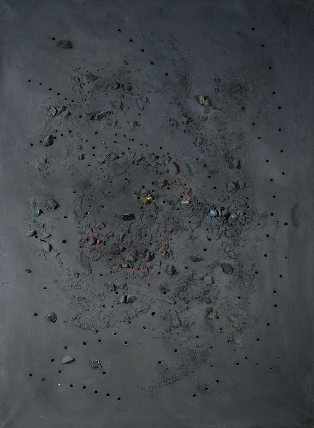
The first painting is the most involved of the three. Part of the “Buchi” (holes) series, it is characterized by a multi-hued dark gray background, colored and clear glass gems, and holes punched into the canvas.

Analysis found that multiple kinds of material were used in the painting, including a variety of pigments, powders, paint extenders and binders. Viridian was founded in the green-tinged areas, while Chrome orange (probably mixed with Chrome yellow) was found in the rust-colored areas. Two different binding agents ders—vinyl acetate and alkyd—were used, one likely to help the paint stick to the canvas, while the other was likely used to keep the gems in place.
The next two paintings are from the “Tagli” (slashes) series and are representative of Fontana’s most iconic works. Concetto Spaziale – Attesa, 1959 features a matte gray background with a single cut slightly off center on the canvas. Fontana would make the slashes in the canvas with a scalpel or Stanley knife. The back of canvas was mounted with black gauze, to filter light and highlight the slashes.

Analysis found evidence of barium, calcium, zinc and sulfur, indicative of white pigments and extenders (substances used to thin and stretch paint). The white pigments were likely mixed with Carbon black to make the distinctive gray color.
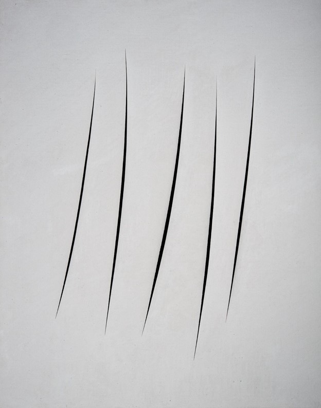
The third painting, Concetto Spaziale – Attese, 1961, has five slashes clustered near the center of the canvas, but set at varying angles and lengths, on a white/light gray background.

The XRF analysis showed titanium, calcium and sulfur, indicative titanium dioxide white pigment. Calcium carbonate was likely used as an extender while polyvinyl acetate was used as a binder.
Understanding and Preserving Fontana’s Legacy
Fontana was known to experiment with different methods in his artwork, which also included sculpture. The spectroscopic analysis of his work proves that to be true, highlighting that the artist used different kinds of pigments, binders and extenders in many of his paintings as he aimed to present just the right color or affect. The data shows that Fontana continued to evolve his methods, even as his career came to a close.
“The combination of a multi-technique approach in material characterization of Lucio Fontana’s artworks resulted fundamental to maximize the information about the artist’s technique: the use of atomic, molecular and vibrational spectroscopies with different probes provided multiple confirmations about Fontana’s choices for pigments and binders, while the contemporary use of portable and laboratory-based experimental techniques was essential to obtain information about surface and stratigraphic distribution of different material on the artworks,” the researchers wrote.
The data provided by the spectroscopic analysis of his works will help museum conservators maintain his work so that it can continue to be enjoyed well into the future.


 Layer It Up: Studying Renaissance Paintings with Multispectral Spectroscopy
  Finding the truth: X-Ray and Art